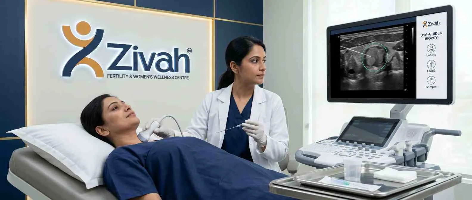

A scan could show that there is something there. It can't tell you what it is. This gap is filled by a USG-guided biopsy, which uses live ultrasound imaging to guide a thin needle to the precise location so that a small sample of tissue may be extracted and evaluated in a laboratory.
It is less intrusive. There is no radiation, major incision, or, in most cases, general anaesthesia involved. The region itself is numbed the whole time you are awake.
In this guide, we will explain why the procedure is recommended, which cancers and organs it can assess, how to prepare, what happens on the day, and when you will receive your findings. Because a scan can create a query, only tissue can answer that.
What Is a USG Guided Biopsy?
This is a test, not a treatment. A small piece of tissue is taken with a thin needle and then sent to a laboratory, and it is seen on a screen during a live ultrasound of the lesion. Ultrasound is safe to repeat and safe in pregnancy, as it employs sound waves rather than ionising radiation, and is therefore the recommended method whenever the lesion is clearly shown on the image.
How Real-Time Ultrasound Guides the Needle
And the radiologist can view the needle tip on-screen the entire time it travels. It’s all about that live view. Small tumours may be targeted with pinpoint accuracy, while avoiding neighbouring blood vessels and organs.
Does a Biopsy Mean I Have Cancer?
No, and here it is worth talking straight. To discover what something is, it is ordered. Not because anything has been proven. A scan can tell you what a lesion is; tissue names it. Many lumps are tested this way and are completely benign.
Why a USG Guided Biopsy Is Advised
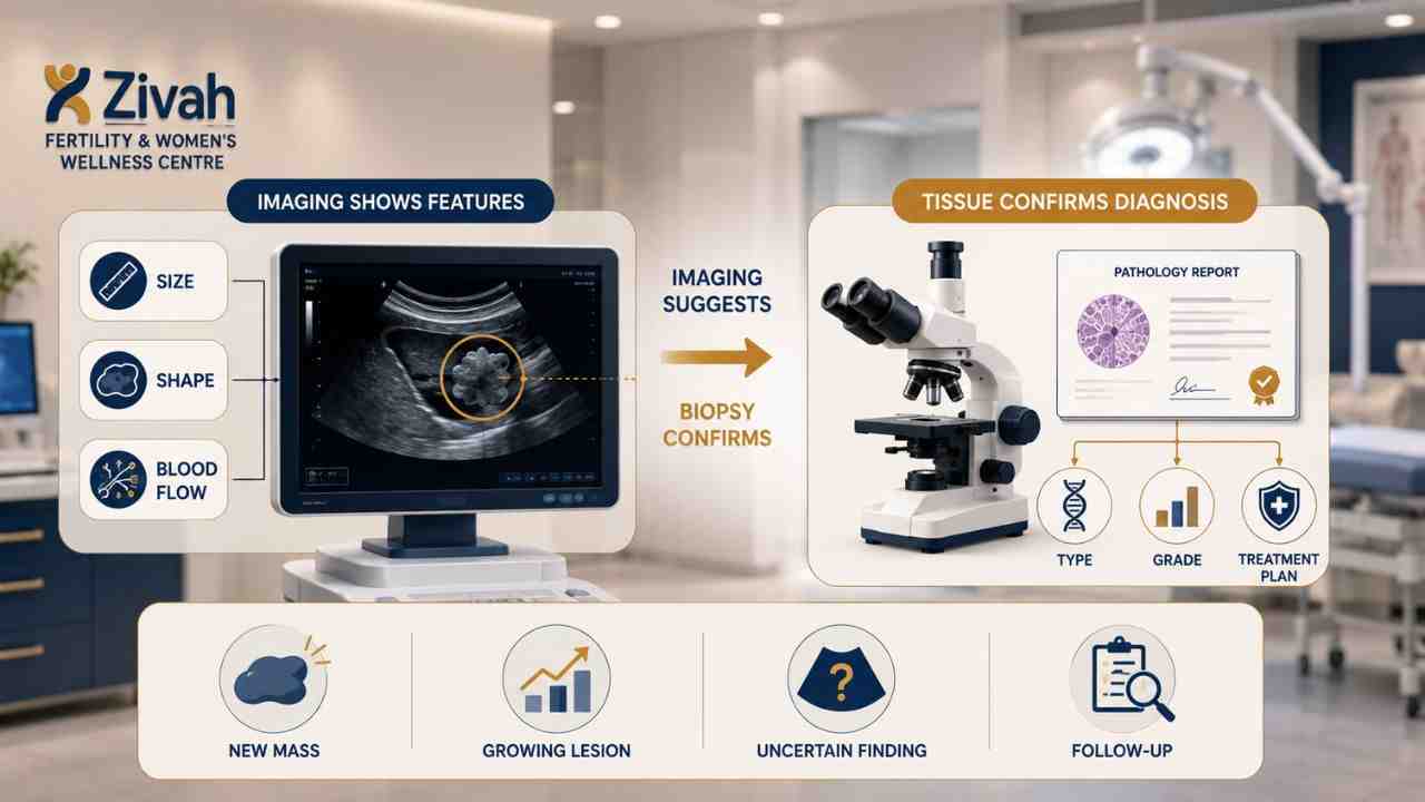
A scan might inform you that a lesion seems irregular, or that it has changed in size since the previous time. What it can't do is tell you what that lesion really is. That can only be answered by examining tissue under the microscope.
When Imaging Alone Cannot Give a Diagnosis
Ultrasound, CT, and MRI are good for defining form, size, boundaries, and blood flow. But description is not diagnosis, imaging may raise suspicion, not resolve it. There is no guesswork involved with ambiguous or borderline features.
You never start treatment just because you suspect anything, since it's the report that tells you the specific type and grade of the tissue, and that tells you whether surgery, chemotherapy, radiation or targeted therapy is the proper course.
Common Reasons Your Doctor Orders a Biopsy
Your doctor could suggest this surgery if:
- There is a new mass or lump on imaging.
- A lesion is developing, irregular or appears aggressive.
- The lesion type is uncertain and cannot be characterised by the imaging.
- It is thought the cancer has spread to another organ.
- Need to examine if past treatment has returned.
When a USG Guided Biopsy Is Recommended
| Clinical Trigger |
Why a Biopsy Is Advised |
What It Confirms |
|---|---|---|
| New lump on imaging |
Imaging cannot identify what the tissue is |
Whether it is benign or malignant |
| Enlarging or irregular lesion |
Growth and irregular borders need explaining |
The nature and behaviour of the lesion |
| Lesion type unclear |
Features fall between benign and malignant |
A definite tissue diagnosis |
| Suspected spread |
Confirms whether disease has reached another organ |
Tissue origin and extent |
| Suspected recurrence |
Distinguishes recurrence from scar or healing change |
Whether disease has returned |
Which Organs and Lumps Can Be Biopsied Under Ultrasound?
The rule is simple: if the scan reveals the lesion clearly, it can usually be sampled in this way. That includes the solid organs of the abdomen and most superficial tumours.
1. Breast Lumps and Axillary Lymph Nodes
This is the most popular usage of all. Core needle is the norm here. They need to grade the breast tissue and analyse it for markers, and the only way to do that is with an intact tissue core. You may also be informed that a little marker clip will be left behind.
This matters to individuals, thus it is important to state clearly: it is routine, you will not feel it, it is safe with MRI, and its only role is to demonstrate precisely where the sample was collected if imaging or surgery follows later.
2. Liver, Kidney and Abdominal Lesions
They sit deeper; therefore, prep is more important. Clotting examinations before a liver or kidney biopsy are more strict, as these organs have a more abundant blood supply.
Subsequently, a small amount of blood in the urine following a kidney biopsy might be normal. You’ll be watched for it, not sent home not knowing.
3. Thyroid Nodules, Neck Nodes and Superficial Lumps
The nodules are frequently collected using a tiny needle (not a core needle), faster, less painful, and sufficient for diagnosis in most situations. Neck nodes are routinely sampled to see whether illness has spread. Lumps near the skin surface are the easiest of all: quick to reach and have the lowest complication rate of any biopsy location.
Types of Needle Biopsy: FNAC, Core Needle and Vacuum-Assisted
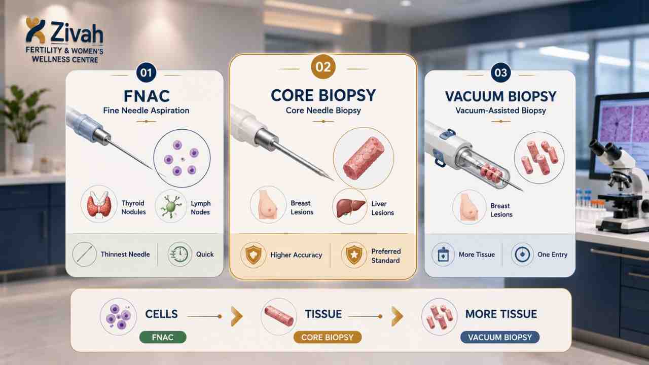
Not every biopsy is done with the same needle. The choice relies on the organ, the size of the lesion and how much detail the pathologist needs to arrive at a solid answer. It is a clinical choice, made for you, not one you have to weigh up yourself.
i) FNAC: Fine Needle Aspiration Cytology
The gentlest one. The thinnest needle pulls out single cells, not a solid section of tissue: fastest, least intrusive and the least unpleasant of the three. It is good for thyroid nodules and lymph nodes. Its drawback is detail. It gathers cells, rather than structure; thus, it may sometimes not grade a tumour or perform the complete battery of laboratory tests.
ii) Core Needle Biopsy: The Tissue Standard
Here, the needle is a little bigger, and it takes a strip of tissue as a whole rather than loose cells. That unbroken structure is what allows a pathologist to evaluate the tissue appropriately and perform marker tests like IHC, which is why it is chosen for breast and liver tissue. Typically, a few tiny samples are obtained in a single visit, which increases accuracy and reduces the chance you'll need to return for a repeat.
iii) Vacuum-Assisted Biopsy: More Tissue, One Insertion
Primarily used in the breast. A mild suction pulls many samples through a single needle entry, so the pathologist receives more tissue, and you get only one puncture.
FNAC vs Core Needle vs Vacuum-Assisted: Sample, Detail and Best Use
| Feature |
FNAC |
Core Needle |
Vacuum-Assisted |
|---|---|---|---|
| Needle size |
Very thin |
Slightly wider |
Wider, vacuum device |
| What's collected |
Cells only |
Intact tissue core |
Several tissue samples |
| Detail provided |
Basic |
Full structure |
Full structure, more volume |
| Grading & markers |
Not always possible |
Yes |
Yes |
| Typical use |
Thyroid, lymph nodes |
Breast, liver |
Breast |
| Discomfort |
Least |
Mild |
Mild |
How a USG Guided Biopsy Is Performed at Zivah
Most of the fear is of the unknown. But here it is, in all its bare skin. You're awake the whole time. The sampling itself is about 15 to 30 minutes, and with the observation after, you should allow a couple of hours at the centre. Not the whole day. It is an out-patient operation, and you go home the same evening. Here’s the step-by-step process:
- You get up and are scanned. You will lie on the table with the area exposed for a breast biopsy, this typically means you will lift your arm. Your radiologist does the scans first to locate the exact area, marks the entrance point, then cleans the skin. None of this hurts. The ultrasound gel is just chilled.
- Only the site is anaesthetised. Let's be straight, this is what everyone asks about. It's local anaesthetic, not general. You're awake, you know what's going on. You feel a little sting when the anaesthetic goes in and then pressure or a pressing sensation, but you should not experience pain. If so, say so.
- The needle is inserted under live imaging. Your radiologist monitors it all on television, guiding the tip to just the right spot. Ultrasound guiding is all about that real-time vision nothing is done blindly. Several little pieces of tissue are removed.
- You'll hear a click. That’s normal. The biopsy equipment takes a forceful mechanical snap each time it samples. It surprises pretty much everybody. Your radiologist will tell you in advance, the sound only signals the sample has come away cleanly.
- Dressing, pressure, done. Firm pressure is used to halt any bleeding, a tiny bandage is put on, and you are carried through to recovery.
Preparing for Your USG Guided Biopsy
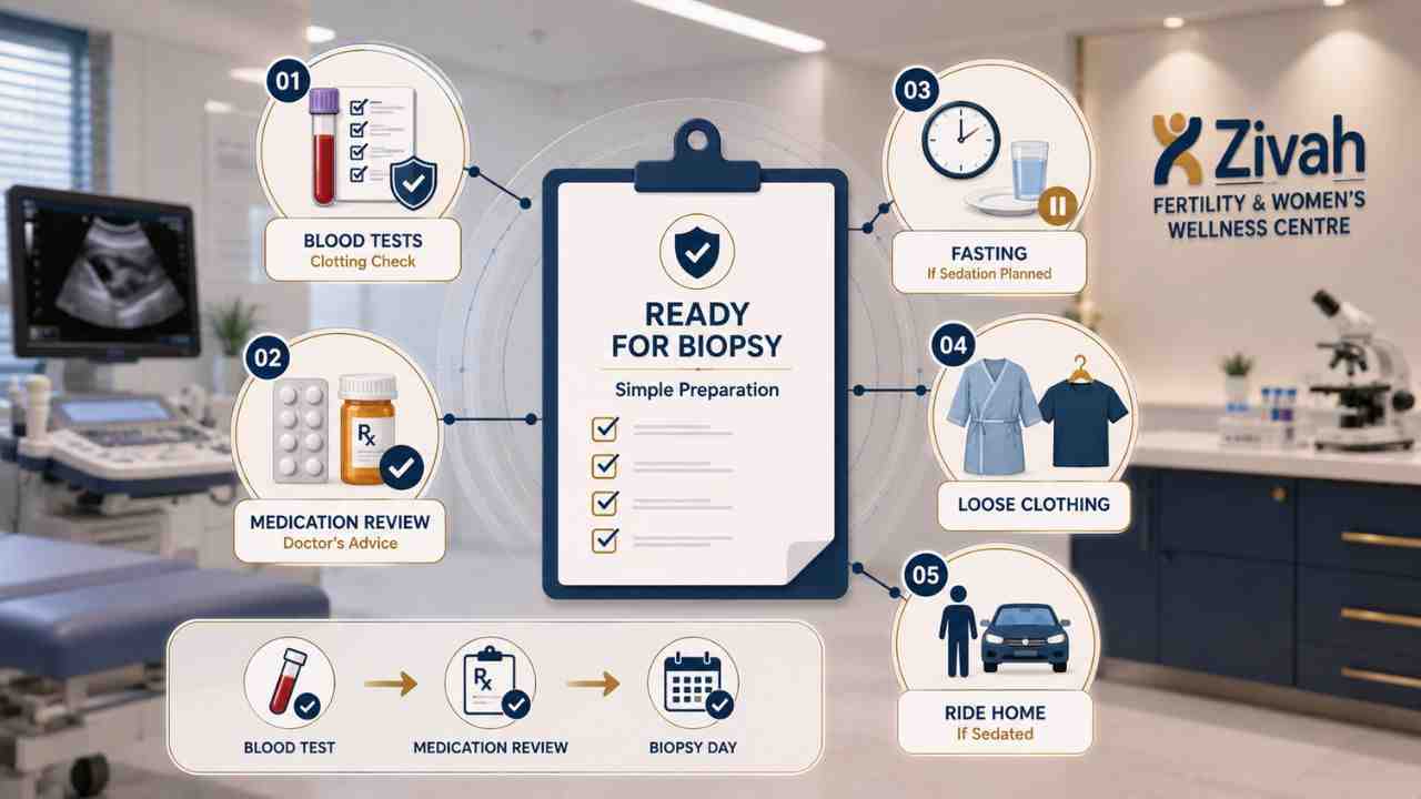
The preparation is mostly about one thing: bleeding risk. The most common reason a biopsy is delayed on the day is an unstopped blood thinner.
Blood Tests and Clotting Checks Before Biopsy
Your team needs to know that your blood clots normally before a needle goes anywhere. This is taken care of by certain standard testing, generally done a few days prior. The examinations are tougher for certain sites; a liver or kidney biopsy is more than a superficial lump.
Medications, Fasting and What to Bring
Tell your doctor about all the medications you are taking, including:
- Blood thinners (warfarin or heparin)
- Aspirin and other comparable medicines
- Anti-inflammatory painkillers, such as ibuprofen
- Supplements that thin the blood more than people know
Some will need to be paused, but only if your doctor says so. Never stop taking a medication on your own. On the day: fasting is only necessary if sedation is planned. Wear loose clothing, be prepared to wear a robe, and arrange transportation home if you are sedated.
Pre-Biopsy Checklist: Action, Timing and Why It Matters
| Preparation |
Action Required |
Why It Matters |
|---|---|---|
| Blood tests |
Done a few days before |
Confirms your blood clots normally |
| Blood thinners |
Pause only if your doctor advises |
Reduces bleeding at the biopsy site |
| Aspirin / painkillers |
Disclose and pause if instructed |
Both increase bleeding risk |
| Supplements |
Tell your doctor what you take |
Some thin the blood unexpectedly |
| Fasting |
Only if sedation is planned |
Keeps sedation safe |
| Allergies |
Disclose before the procedure |
Especially to anaesthetic |
| Escort home |
Arrange if being sedated |
You cannot drive after sedation |
After the Biopsy: Recovery Time and Aftercare
Recovery from an ultrasound-guided biopsy is fast and easy. No cut to heal, only a little puncture site that has to be protected while it heals. Most people are back to their normal schedule the next day. Knowing what is normal and what is not is important.
Observation, Dressing and Same-Day Discharge
Samples are taken from the tissue and pressure is applied firmly with a tiny dressing. If the biopsy were from a deep organ (such as the liver or kidney), you would be monitored longer before being discharged than if it was from a surface tumour. Same-day routine discharge.
Wound Care, Returning to Work and Restarting Blood Thinners
Keep the dressing dry and clean, relax for about 24 hours, and avoid heavy lifting for a few days. Usually, you're OK to go back to a desk job the next morning. Don't drive if you were sedated.
If you stop a blood thinner, wait until your team says the site has sealed before beginning again. Never resume on your own. Call us immediately if the bleeding seeps through the dressing, pain increases rather than improves, you develop a fever, have redness or pus at the site, find blood in your urine after a kidney biopsy, or have shortness of breath or chest discomfort.
Biopsy Results: How Long the Report Takes and What It Tells You
Let's be honest here: the waiting is the worst part of the whole procedure. Ultrasound-guided biopsy itself takes half an hour. But it's the days after that folks have a problem with. So it's good to have an idea of what's occurring with your tissue sample while you wait.
Biopsy Report Time: Standard Turnaround vs Special Testing
The pathologist looks at the tissue in your sample using a microscope. It normally takes a few days to get a simple biopsy report back. When further testing is required, it might take longer for biopsy findings, immunohistochemistry (IHC) for identifying tumour markers or molecular and genetic testing for targeted treatment. The bit to hold on to: longer delay = more comprehensive testing, not worse news. People think delay is a bad outcome. It doesn't.
What Your Biopsy Report Shows and What If It's Inconclusive
The report will establish whether the tissue is benign or cancerous. If it is malignant, it will specify the type of tumour, the grade and any relevant symptoms, the information that will guide your treatment approach. Biopsy report should not be viewed in isolation.
Your doctor will review it with you. Sometimes the sample is insufficient to provide a definitive diagnosis. An inconclusive biopsy doesn't necessarily mean something is wrong; it generally requires a repeat or surgical biopsy to obtain additional tissue.
Is a USG Guided Biopsy Safe? Risks and Side Effects
It's a standard, low-risk process. It's a procedure. Serious consequences are rare, but you owe it to yourself to hear the real story before you say yes to anything.
Common Side Effects of USG Guided Biopsy vs Rare Complications
Common side effects are mild. Soreness at the spot, some bruising, a little blood from the skin; it all settles within days, and basic pain treatment is generally adequate.
Major consequences are quite rare: major internal bleeding (particularly important after a liver or kidney biopsy), infection at the site, or harm to a neighbouring structure. This live ultrasound imaging is precisely what makes them uncommon; the needle is never advanced blindly.
Who Is at Higher Risk at USG Guided Biopsy
First and foremost is bleeding risk. If you have a bleeding condition, are on blood thinners, or need a deep-organ biopsy, such as the liver, for example, which is highly vascularized, you may need special care. Sometimes you delay a biopsy. If the clotting can't be safely corrected, or the lesion can't be accessed without going through something it shouldn't.
Side Effects vs Complications: What's Common, Rare, and Needs Attention
| Category |
What It Is |
How Common |
What to Do |
|---|---|---|---|
| Side effect |
Soreness at the biopsy site |
Common |
Simple pain relief; settles in days |
| Side effect |
Bruising or minor skin bleeding |
Common |
Keep dressing clean and dry |
| Complication |
Bleeding under the skin (haematoma) |
Uncommon |
Apply pressure; contact your team |
| Complication |
Infection at the site |
Rare |
Redness, pus or fever, call us |
| Complication |
Significant internal bleeding |
Rare |
Worsening pain, dizziness, urgent care |
| Complication |
Injury to a nearby structure |
Very rare |
Managed by your clinical team |
USG Guided Biopsy vs Other Biopsy Methods
Your doctor’s technique is not a random choice. The appearance will depend on the position and depth of the lesion as well as its conspicuousness for each modality of imaging.
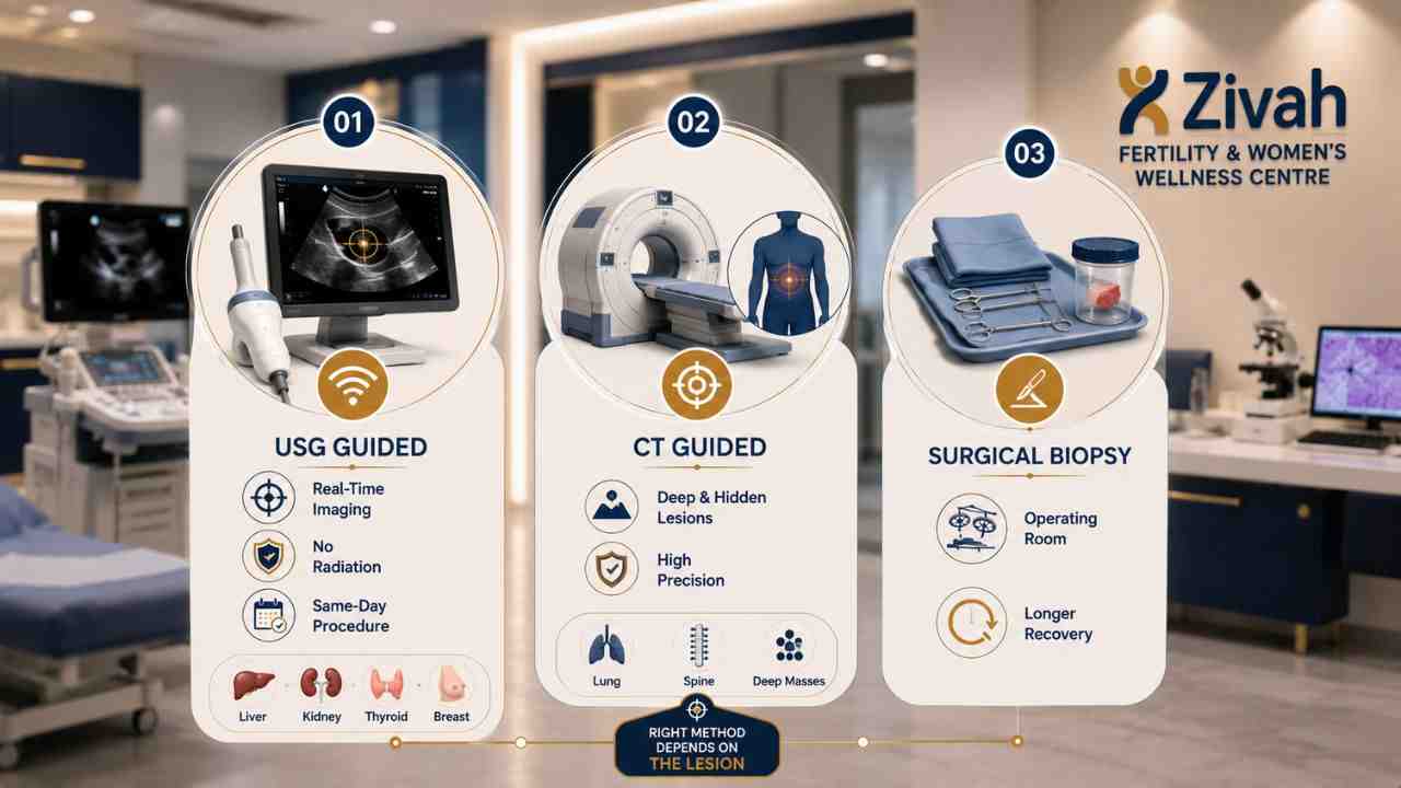
Ultrasound vs CT Guidance: Which Is Used When
When the lesion is plainly visible, liver, kidney, thyroid, breast, and most superficial tumours require ultrasound guidance. It lets you see the needle in real time, without radiation.For deep or concealed lesions such as lung nodules, retroperitoneal masses, or spinal lesions, CT-guided biopsy takes over, mapping a safe path around organs and veins.
Needle Biopsy vs Surgical Biopsy: Accuracy and Recovery
Surgical biopsy means removing part or all of the lump through an incision. This is generally done under general anaesthetic and involves sutures with a prolonged recovery.
An image-guided needle biopsy makes the same diagnosis with a puncture a few millimetres wide, no general anaesthetic, no scarring, and same-day discharge. High precision is ensured by live needle visualisation and repeated sampling.
USG-Guided vs CT-Guided vs Surgical Biopsy: Comparison
| Feature |
USG-Guided |
CT-Guided |
Surgical |
|---|---|---|---|
| Imaging used |
Live ultrasound |
CT cross-sections |
None during surgery |
| Radiation |
None |
Yes |
None |
| Anaesthesia |
Local |
Local, sometimes sedation |
Usually general |
| Incision |
Needle puncture |
Needle puncture |
Surgical cut, stitches |
| Best for |
Breast, thyroid, liver, kidney, superficial lumps |
Lung, spine, deep abdominal lesions |
When needle sampling is insufficient |
| Recovery |
About a day |
About a day |
Several days to weeks |
| Discharge |
Same day |
Same day |
Same day or overnight |
Why Choose Zivah for Your USG Guided Biopsy
A biopsy's quality depends on the person doing the biopsy and the lab examining the tissue. Both are worth asking about, wherever you go for it. You see, a scan might ask a question. The tissue can answer it alone.
A USG-guided biopsy is quick, minimally invasive and done in one visit: a little needle, live imaging and a sample that ultimately tells you what you are dealing with. Whatever the report says will determine your therapy. It's what takes confusion out and puts a strategy in place.
Understanding Biopsy Costs and Insurance
There isn't a set price for an ultrasound-guided biopsy. depends on the organ and depth, the type of needle, whether anaesthesia is needed, and which pathological tests are necessary; IHC and molecular investigations add to it.
Call our staff to get a true quote and find out what your insurance will cover. If a scan has revealed something that requires explanation, schedule a consultation. We will explain what is involved before anything is booked.