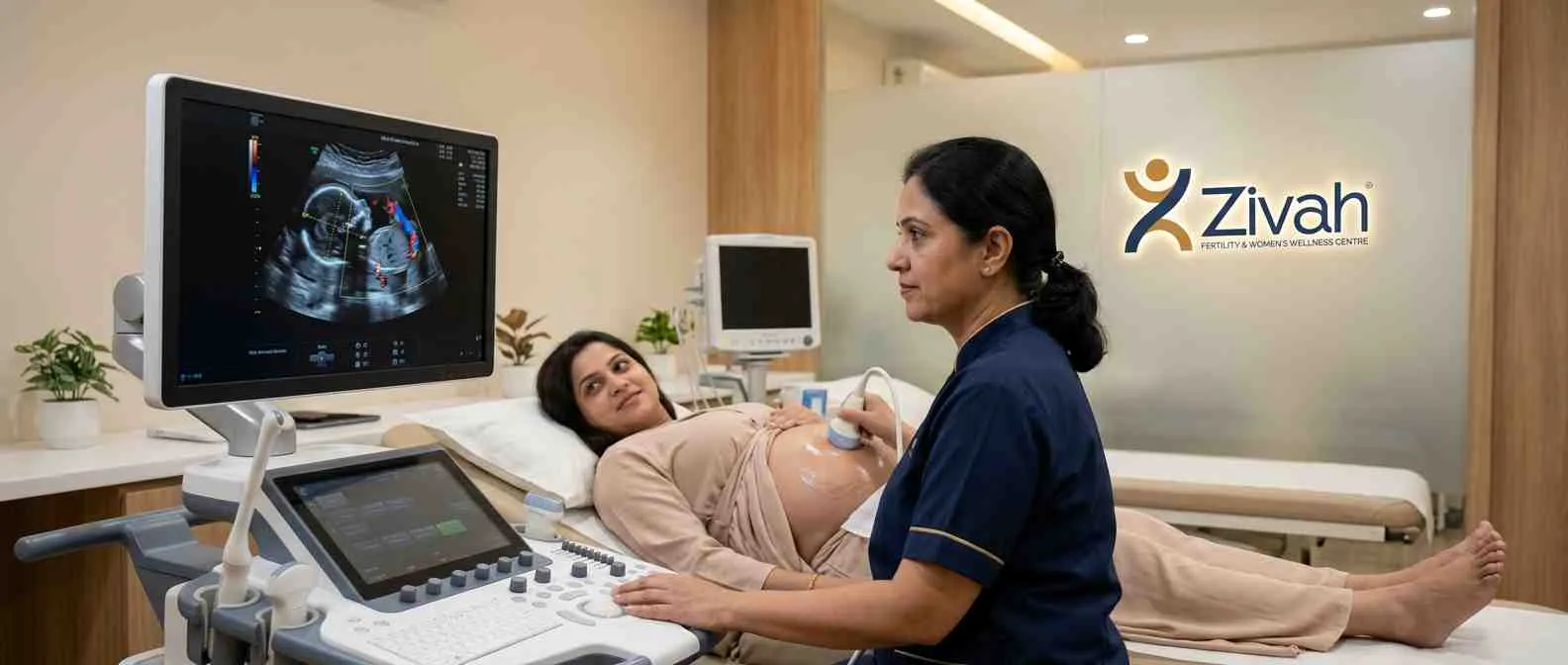
A USG biometry, well-being, and Doppler scan combines three important tests in one painless ultrasound. The biometry part looks at your baby’s growth, the fetal wellness scan examines how well your baby is doing, and the fetal Doppler scan checks blood flow between you, the placenta and your baby. This pregnancy ultrasound, usually done later in pregnancy, gives your specialist a good view of how things are progressing.
On this page, you will discover what each part of the scan checks, why and when it is done, how to prepare and what the results signify. It’s so useful because one fetal growth scan gives you three different checks, and each check answers a different question about how your baby is growing and healthy.
Important: Our Legal Compliance Commitment - This scan is just to check your baby’s growth, welfare and blood flow. In accordance with the PCPNDT Act, 1994, Zivah does not ascertain or divulge the sex of the infant under any circumstances.
What Is a Fetal Growth, Wellbeing & Doppler Scan?
In the later weeks of your pregnancy, one image isn’t always enough to know if your baby is doing okay. That’s why this combination scan is so helpful, one simple ultrasound that does three duties at once. A fetal biometry scan checks how your baby is growing. A well-being check looks for evidence that your infant is doing well.
A fetal Doppler scan checks the blood flow between you and your baby. The growth and Doppler scan combine everything into one visit instead of three separate consultations. It’s non-invasive, typically done in the third trimester and allows your specialist to get the full picture in one visit.
Biometry, Wellbeing and Doppler: Three Checks in One Scan
Each section solves a separate question:
- USG biometry - Is my baby developing normally?
- Fetal wellness ultrasound - How is my baby doing now?
- Fetal blood flow scan - Is the circulation maintaining the fetus properly?
Taken together, this comprehensive scan of fetal wellness gives a more complete picture than any one check can.
How a Pregnancy Ultrasound Assesses Your Baby Safely
The scan uses harmless sound waves, a reliable technology used throughout your pregnancy. These waves are sent through the belly during a prenatal ultrasound, and the echoes that bounce back are analysed. Colour Doppler adds a view of blood flow.
Ultrasound is a safe way to image you and your baby throughout pregnancy because it does not use radiation, and nothing is put into your body. What each section of the scan examines.
What Each Part of the Scan Checks
| Component |
What It Measures |
Why It Matters |
|---|---|---|
| Biometry |
Head, abdomen, femur size; estimated weight |
Tracks growth against gestational age |
| Wellbeing |
Amniotic fluid, movement, heart rate |
Confirms the baby is thriving |
| Doppler |
Blood flow in cord, brain, uterus |
Detects circulation problems |
Fetal Biometry: Measuring Your Baby's Growth (BPD, HC, AC, FL)
The scan's initial function is to measure your baby. This is fetal biometry, a series of meticulous measurements that reveal how your baby is growing compared to what is expected for this time of pregnancy.Your specialist will take many measurements of fetal biometry and use a combination of them rather than a single number. Together, they answer the key question driving every newborn growth scan: Is your baby growing on track?
The Four Key Biometric Measurements: BPD, HC, AC & FL
The typical ultrasound for the growth of the fetus includes four measurements:
- BPD (biparietal diameter) - the width of the baby's head from side to side
- HC (head circumference) - the distance around the head
- AC (abdominal circumference) - the measurement around the tummy, an important clue to growth and nutrition
- FL (femur length) - the thigh bone length, the longest bone in the body
Each is taken from a specific point of view to ensure accurate readings.
Estimated Fetal Weight (EFW) and Growth Percentiles
Once those four measures are in, they are put together to provide an estimated fetal weight, which is what your baby likely weighs right now. This is then represented as a percentile on a growth chart, where fetal weight estimation becomes particularly useful.
The percentile shows how your baby compares to others of the same age and helps to flag if a baby is measuring "small for dates" or "large for dates." It's an easy, reassuring approach to observe growth over time, tracked throughout scans.
The Four Fetal Biometry Measurements Explained
| Measurement |
What It Assesses |
Abbreviation |
|---|---|---|
| Biparietal diameter |
Head width |
BPD |
| Head circumference |
Head size |
HC |
| Abdominal circumference |
Tummy size (growth/nutrition) |
AC |
| Femur length |
Thigh bone length |
FL |
Fetal Wellbeing Assessment: Amniotic Fluid, Movement & Heartbeat
Measuring your baby’s size gives you part of the story, not the whole story. A baby can be the correct size and still need to be monitored to see how they are doing day to day. This is what the fetal wellbeing scan is for, checking for the signals that your baby is happy and thriving in the tummy.
This phase of the fetal assessment scan looks at a few critical signs beyond growth, the fluid around the baby, how active they are, the heartbeat, and the placenta that feeds them.
Amniotic Fluid Index (AFI) and What It Indicates
The fluid that surrounds the baby might tell you a lot about its condition. Your specialist checks the pockets of fluid in the womb by measuring the amniotic fluid during the amniotic fluid evaluation and combines them into one result, the amniotic fluid index or AFI.
A healthy AFI means the infant is properly supported. This AFI scan is a standard aspect of the evaluation, as too little or too much can be a warning flag to watch out for.
Fetal Movement, Heart Rate and Placental Check
Good signals that your baby is doing well are movement and heartbeat. As part of the examination of foetal mobility, your specialist will watch how your baby moves during the scan. In contrast, the ultrasonography of the fetal heart rate shows that the heartbeat is constant and strong.
The placenta is checked too, including its position and, where needed, a placental Doppler scan to see that it's delivering blood and nutrients as it should.
Fetal Doppler Scan: Assessing Your Baby's Blood Flow
Growth and well-being tell you what your baby looks like today, but a fetal Doppler scan shows something that you can’t see in a measurement: how the blood is actually moving.
Your specialist will do pregnancy Doppler ultrasounds to examine the flow between you, the placenta and your baby to make sure oxygen and nutrients are being delivered effectively.
Umbilical and Uterine Artery Doppler
The first vessels to be examined are the lifelines of the pregnancy. Umbilical artery Doppler looks at the flow of the cord, which helps tell how quickly blood is moving through the placenta, a big signal about how well it’s operating.
The uterine artery Doppler is a look at the supply from your side, the maternal-fetal circulation feeding the placenta. They warn of early signs of placental problems. Blood flow to the baby’s brain matters too.
MCA, Cerebroplacental Ratio (CPR) and Ductus Venosus
Use the middle cerebral artery Doppler, which measures blood flow to your baby’s brain. CPR combines the umbilical and brain values into a single ratio, providing a clearer overall picture of fetal circulation.
During more intensive monitoring, the ductus venosus, a vessel close to the baby’s heart, is examined as a safety flag in late pregnancy. For accuracy, Zivah specialists measure the MCA at the precise 0-15° angle. Our blog explains the deeper meaning of each measurement.
Color, Pulsed and Power Doppler Methods
The readings are obtained using different Doppler modes. Colour Doppler in pregnancy is a technique for visualising blood flow. It adds direction and speed to the image so you can see it easily at a glance.
Pulsed wave Doppler monitors flow in a single specific location. Power Doppler detects the tiniest signals. Together, this obstetric colour Doppler delivers a complete view of your baby’s circulation.
Key Doppler Vessels and What They Assess
| Vessel |
What It Reflects |
Why It's Checked |
|---|---|---|
| Umbilical artery (UA) |
Placental resistance |
Core IUGR marker |
| Uterine artery (UtA) |
Maternal–placental flow |
Pre-eclampsia / growth risk |
| Middle cerebral artery (MCA) |
Brain blood flow |
Detects "brain-sparing" |
| Ductus venosus (DV) |
Fetal heart strain |
Late-stage IUGR safety marker |
Why and When a Third-Trimester Growth & Doppler Scan Is Done
This scan is most typically part of looking after you in the final stages of pregnancy. If you’ve ever wondered why a fetal doppler scan is done, it’s usually for one of two reasons: routine growth tracking or to keep a closer eye when something needs monitoring. It is a third-trimester scan, providing your specialist with up-to-date information when growth and blood flow are most important.
High-Risk Pregnancy Monitoring and Growth Restriction (IUGR)
Doppler obviously has a place in high-risk pregnancies. A Doppler for high-risk pregnancies can pick up early warning signs of problems and make a real difference to outcomes for illnesses such as high blood pressure, diabetes or a baby that is smaller than expected.
It is especially useful in cases of suspected fetal growth restriction (IUGR), as early detection of abnormalities allows appropriate therapy. At Zivah, we recommend Doppler when it provides true therapeutic value, is evidence-driven, and not routine.
Routine Third-Trimester Growth Monitoring
For many pregnancies, the scan is simpler, a steady third-trimester growth scan to confirm your baby is growing well over time. This is a fetal development scan based on size and growth rather than precise anatomy, that detailed structural check is done earlier, in the Anomaly Scan (TIFFA). If you’re earlier on in pregnancy, the NT Scan and Early Pregnancy Scan are the places to start.
How the Fetal Doppler Examination Is Done and How to Prepare
The good news: This scan doesn't require much of you. A fetal Doppler is a gentle, non-invasive test that is done from the outside of your abdomen, just like any antenatal ultrasound scan you have had before.
No needles, nothing is put in you, it's completely painless. And to address a common concern: ultrasonography is safe during pregnancy, utilises no radiation and has no known danger to your baby.
Preparing for Your Growth and Doppler Scan
You don't need to do much to prepare for a pregnancy ultrasound: Wear loose two-piece apparel so your abdomen is easily accessible You can normally eat and drink; no fasting is required If your expert wants them to bring any previous scan reports That’s about all the prep required for a regular pregnancy ultrasound.
What Happens During the Scan
Once you're in place, it's really straightforward. You lie down, and a little gel is spread on your stomach, and a specialist slowly moves a transducer over it. During this obstetric ultrasound, they capture the growth measurements, well-being checks, and Doppler blood flow in one sitting, fetal-medicine-led, painless, and usually done in 20 to 40 minutes.
Your Growth & Doppler Scan: Step by Step
| Stage |
What Happens |
Time |
|---|---|---|
| Before |
Minimal prep; lie down |
~5 min |
| During |
Biometry + wellbeing + Doppler captured |
20–40 min |
| After |
Resume normal activity |
Immediate |
Understanding Your Growth & Doppler Results: Normal vs IUGR
First, something reassuring to remember: the notes below are general context to help you understand your scan, not a diagnosis. Your growth scan results are always reviewed by a specialist in fetal medicine who considers them in the context of your whole pregnancy history before anything is decided. Most scans come with positive news. If your baby’s development and blood flow are where they should be, that’s a good sign that things are on track.
Normal vs Abnormal Growth and Doppler Findings
If the test is normal, it suggests your baby is growing on target and that the blood flow appears healthy. If anything is outside the predicted range, it does not inherently signify trouble; it only suggests it is worth taking a closer look. For instance, if the infant is below the predicted percentile, your specialist may look out for fetal growth restriction (IUGR).
Similarly, abnormal Doppler in pregnancy, such as increased resistance in the cord, can suggest the placenta is working harder than it needs to. In all circumstances, the Doppler scan results lead to the next step, usually closer monitoring, not an alarm.
Those are merely high-level pointers. You can read all about how to interpret the actual numbers, pulsatility index, CPR and IUGR staging in our growth and Doppler blog guide.
Scan Findings and What They May Indicate (Specialist-Interpreted)
| Finding |
What It May Suggest |
Typical Next Step |
|---|---|---|
| Growth below expected percentile |
Possible IUGR / SGA |
Closer surveillance |
| Raised umbilical artery resistance |
Placental insufficiency |
Specialist review |
| Reduced amniotic fluid (low AFI) |
Wellbeing concern |
Repeat / monitoring scan |
Findings are interpreted by a fetal-medicine specialist in your full clinical context; they are not a diagnosis on their own.
Fetal Growth & Doppler Scans at Zivah
At Zivah, your fetal wellbeing scan will be done by a fetal-medicine specialist and is meant to provide you with a complete picture in one visit. We combine biometry, wellbeing and Doppler in one complete fetal wellbeing scan, to assess your baby's growth, the indicators they're thriving and the maternal-fetal blood flow, all in one session.
The distinction is in the details. Our specialists check all vessels including the ductus venosus if necessary and measure the MCA at the exact angle for reliable values. And we perform careful scans, suggesting Doppler where it truly adds clinical value, never as a standard addition.
From Scan to Specialist Review and Care Plan
After your fetal biometry and Doppler scan, your data are reviewed by a specialist who explains what they mean and what happens next, reassurance or closer pregnancy growth monitoring.
Every pregnancy is different; thus, the cost is discussed during your appointment.This scan is for growth, wellbeing, and blood flow solely as per the PCPNDT Act; Zivah does not determine or reveal your baby's sex. Watchful eyes for a growing infant. Book your scan with Zivah today.