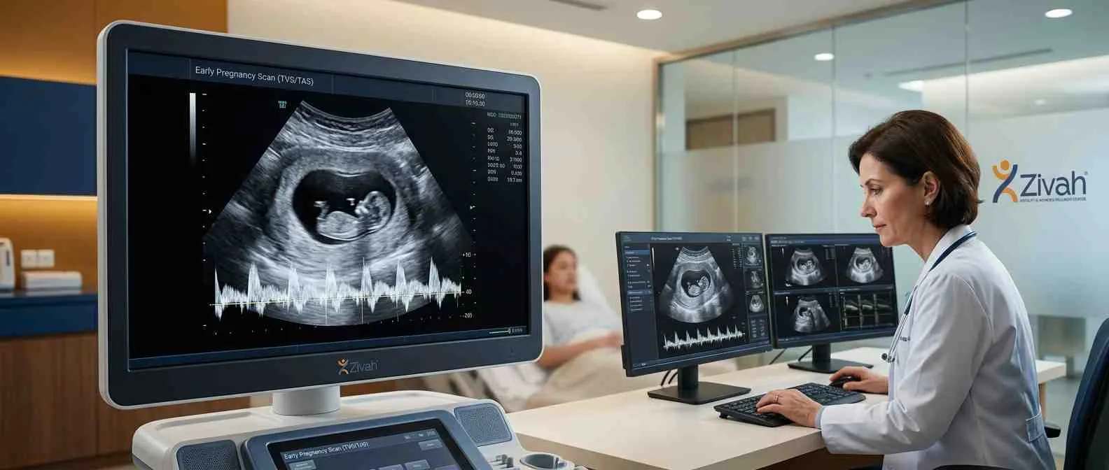
The first ultrasound in pregnancy is known as an early pregnancy scan. It is usually done in the first trimester to confirm pregnancy, check the location, get an estimation of your due date and check early viability. It can be performed by a transvaginal scan (TVS, internal) or a transabdominal scan (TAS, external).
This is your first detailed look at your pregnancy to ensure that it’s developing in the proper place and at the right rate. This page explains what the early pregnancy scan checks, when it happens, and how the two types of early pregnancy scan, transvaginal (TVS) and transabdominal (TAS) scan, work. Going for your first ultrasound during pregnancy? Want to know more about sonography in early pregnancy? Here’s everything you need to know before your visit.
What Is an Early Pregnancy Scan?
An early pregnancy scan, also called a first-trimester ultrasound, is used to confirm your pregnancy, locate it, calculate your due date, and assess early viability. In other words, it's the first detailed check that your pregnancy is in the right place and developing as expected.
This can be done in two ways: as a transvaginal or transabdominal scan. Your prescription could say "TVS": medically, TVS is the short form for transvaginal sonography (also written as TVUS), which is performed by inserting the probe in the vagina. Both are standard procedures in early pregnancy sonography, and your doctor will take the most appropriate one according to your weeks of pregnancy.
What Is a Transvaginal Scan (TVS)?
A transvaginal ultrasound is performed internally. In early pregnancy, a slim, covered, lubricated probe is softly inserted into the vagina to get a close, clear picture of your uterus.
The first few pictures from a TVS sonography are clearer as the probe is nearer to the uterus. A scan done from the abdomen is not as clear. For this reason, a TVS sonogram is also known as an internal sonogram or vaginal sonogram (you may see it printed as USG pelvic TVS).
What Is a Transabdominal Scan (TAS)?
The one which is performed externally is called transabdominal ultrasonography (TAS). A transducer is moved across your lower abdomen, and images appear on a screen. A full bladder is usually required for clarity, and this is desired from the latter part of the first trimester onwards. For example, a transabdominal ultrasonography at 9 or 10 weeks.

Early Pregnancy Scan at a Glance
| Parameter |
Detail |
|---|---|
| Purpose |
Confirm, locate, date & check viability |
| Methods |
TVS (internal) or TAS (external) |
| Best time |
First trimester (~6–10 wk) |
| Duration |
TVS ~5-10 mins; TAS ~10-15 mins |
| Preparation |
TVS: empty bladder; TAS: full bladder |
| Report |
Usually same day |
TVS vs TAS: Is the Early Pregnancy Scan Internal or External?
So, is the first ultrasound internal or external? That depends upon how early you are. In the first trimester, the scan is generally performed internally (TVS), as it provides the best view of this early stage of pregnancy.
The difference is easy. A transvaginal scan (TVS) is an internal scan; the probe is near your uterus. An external transabdominal scan (TAS) is performed over the abdomen. This is why TVS is the best practice and the gold standard for identifying issues such as ectopic pregnancy.
In early pregnancy, an internal ultrasonography detects the gestational sac and heartbeat sooner and more correctly. As you move through your pregnancy, the trans abdominal scan is the usual.
So if you are wondering if the 8-week or 9-week ultrasound will be internal or external, it can be either, but the doctors generally switch to abdominal scans from the later first trimester, but TVS may be performed later in pregnancy if required.
TVS vs TAS Compared
| Feature |
TVS (internal) |
TAS (external) |
|---|---|---|
| Route |
Probe in vagina |
Over abdomen |
| Best stage |
~4–10 wk |
~9 wk onward |
| Bladder |
Empty |
Full |
| Image clarity in early pregnancy |
High |
Low |
Why Is TVS Preferred in Early Pregnancy?
The probe is positioned considerably closer to your uterus, so the images are clearer and the heartbeat can be detected earlier. That's why the 5, 6, 7, or 8-week ultrasound will show more with a transvaginal scan than with an abdominal scan. TVS is more reliable in the presence of a tilted (retroverted) uterus or a broader body type, which can make abdominal imaging more difficult.
When Is an Early Pregnancy Scan Done?
An early pregnancy scan is normally performed between 6 and 10 weeks of your pregnancy. This is the window when there is enough to view and when the scan delivers the most reliable information on location, date and early viability.
When can a sonogram detect pregnancy? The gestational sac can be seen from around 4 to 5 weeks on a transvaginal ultrasound, and the heartbeat is normally seen from around 5.5 to 6 weeks.
It's important to be realistic about the timing, especially if you're wondering if 4 weeks is too soon for an ultrasound. There is little to visualise yet at 1-3 weeks. Very early on, there is little to show. Even an ultrasound at 4 weeks gestation, or 4 weeks 6 days, may only show the early chances of pregnancy.
Can an Ultrasound Detect Pregnancy at 4-6 Weeks?
Yes, but timing is everything. A transvaginal ultrasound can detect a pregnancy at the point when the gestational sac is first seen ( approximately 4 to 5 weeks ). So if you're wondering if you can see pregnancy on ultrasound at 4 weeks, the answer is yes, but you may only see a little sac, not a baby.
The heartbeat is a little late. By roughly 6 weeks, a transvaginal ultrasound can frequently provide a sharper picture, including early heart activity. So, at 4 weeks, you can expect to see very little; however, at 6 weeks, there is usually much more confidence.
How Is an Early Pregnancy Scan Done?
Thinking how a transvaginal ultrasound or how the scan works? It's a basic process, and the steps will depend on the approach your doctor uses.
For a trans vaginal scan (TVS):
- You'll normally be asked to empty your bladder first.
- You are lying comfortably on the examining table.
- A thin probe, covered with a sheath and lubricating lubricant, is placed into the vagina.
- The probe will provide you with clear pictures of your uterus on a screen, and if you prefer, you can have a chaperone there too.

For transabdominal scan (TAS):
- They apply a clear gel to your lower abdomen.
- The images on the screen are taken by a little handheld device called a transducer, which is moved about on your belly.
That's the whole ultrasound process for pregnancy, easy-peasy, and not painful, just the pressure.
How Long Does a Transvaginal Ultrasound Take?
A transvaginal ultrasound takes roughly 5 to 10 minutes. It's a brief, basic internal ultrasound, and you can be right back to your normal day.
Is the Early Pregnancy Scan Safe?
Yes, the early prenatal ultrasound is quite safe. Ultrasound uses sound waves, not radiation, so there's no known risk to you or the baby. A TVS does not raise the risk of miscarriage, and vaginal bleeding will not influence the scan or its accuracy.
Does a Transvaginal Ultrasound Hurt?
A transvaginal ultrasound should not be painful. Most women say it is simply a little pressure, as the probe is thin and gently placed. If you feel anxious, you can ask for a chaperone, ask to place the probe yourself or choose the abdominal scan. Light spotting or cramps for a short while after is usual and not harmful.
How to Prepare for an Early Pregnancy Scan
It’s easy to prepare for an early pregnancy scan, and what you’ll need to do depends on the procedure. The only difference is your bladder. You’ll need an empty bladder for a transvaginal ultrasound (TVS), so it helps to use the restroom right before.
The reverse is true for a transabdominal ultrasound (TAS), you want a full bladder, which normally means drinking around 500 ml of water an hour before your visit. No fasting needed either way. So if you’re thinking "Can you eat before a transvaginal ultrasound", the answer is yes, eat normally, and you can drink water before the exam too.
Do You Need a Full Bladder?
This is where most people do it wrong. In a transabdominal ultrasound (TAS), you require a full bladder, as this pushes the womb up into view more clearly. But for a transvaginal ultrasound (TVS), you want an empty bladder because a full bladder might actually interfere with crisp images.
Can you have the scan if you are experiencing bleeding during pregnancy?
Yes, you can have the scan if you are bleeding in pregnancy, often in fact it is when it is needed. A transvaginal ultrasound is a safe, gentle way to find out the cause of the bleeding, check your baby’s heartbeat and rule out issues like ectopic pregnancy or early miscarriage.
Scan Preparation: TVS vs TAS
| Aspect |
TVS |
TAS |
|---|---|---|
| Bladder |
Empty |
Full |
| Eating |
Normal |
Normal |
| Having bleeding |
Fine |
Fine |
What Can Be Seen on an Early Pregnancy Scan?
What you see on an early pregnancy scan depends largely on how far gone you are; the picture changes week by week. In the first weeks, the uterus pregnancy ultrasound shows the gestational sac, the first clear evidence on a sonogram in early pregnancy. Shortly thereafter, the yolk sac and fetal pole are seen, then the embryo and, a little later, the heartbeat.
So if you're wondering what you can see on an ultrasound at 4 weeks, the answer is minimal, generally just a little sac. As things go forward, an embryo ultrasound also allows your sonologist to estimate the crown-rump length (CRL) for correct dates. Two more things are important at your pregnancy's first scan:
- To verify that the pregnancy is inside the uterus (not ectopic)
- To verify whether you are carrying one or multiple babies
There is hardly any movement thus early, so the focus is on these fundamental structures.
What the Scan Shows by Week
| Weeks |
Typically visible (TVS) |
What it confirms |
|---|---|---|
| ~4 - 5 wk |
Gestational sac |
Early pregnancy |
| ~5 - 6 wk |
Yolk sac, fetal pole |
Developing pregnancy |
| ~6 wk+ |
Fetal heartbeat |
Viability |
| ~7 - 8 wk+ |
Clearer embryo, CRL |
Growth & dating |

This is for indication purposes only; visibility differs from pregnancy to pregnancy and should be interpreted by your provider. This is not a timeline of self-assessment.
Detecting the Fetal Heartbeat and Pregnancy Location
One of the most obvious early signals that a pregnancy is progressing is the fetal heartbeat, which may be seen from about 5.5 to 6 weeks with a transvaginal scan. Cardiac activity is frequently well established by the time of a 7-week trans abdominal ultrasound. And just as importantly, the scan confirms where the pregnancy is located.
A transvaginal ultrasound can confirm pregnancy and determine where the fetus is located. One of the first things to do is to make sure it's not an ectopic ( pregnancy outside the uterus ).
Early Scan Report Terminology Decoder
| Term |
Stands for |
Plain-English meaning |
|---|---|---|
| GS |
Gestational Sac |
Fluid sac around baby |
| YS |
Yolk Sac |
Early nourishment structure |
| CRL |
Crown–Rump Length |
Baby's length, head to bottom |
| FHR |
Fetal Heart Rate |
Heartbeat seen on scan |
| MSD |
Mean Sac Diameter |
Size of the gestational sac |
Understanding Your Early Pregnancy Scan Results
An early pregnancy ultrasound that reassures will usually confirm three things:
- The pregnancy is uterine (not ectopic)
- Heartbeat present (at roughly 6 weeks)
- The dating works the timeline expected
Together, these confirm that the pregnancy is doing nicely. Want to learn how to read a transvaginal ultrasound or a pregnancy ultrasound report? Your sonologist does not work on a single figure; they look at the gestational sac, yolk sac, foetal pole, heartbeat, and measurements such as CRL, then note them all down. A normal ultrasound of the uterus simply suggests these things are present and developing as they should for your stage.
What If the Scan Shows a Problem?

Remember that the results of pregnancy scans are not read in isolation but are considered within the context of your entire history and dates. Don’t jump to the worst conclusion when you encounter the unexpected. A seemingly incorrect date measurement is often a question of revised dating, and a second scan in 1 to 2 weeks makes it clear. The early scan can also pick up on valid issues such as:
- Ectopic pregnancy: implantation outside the uterus
- Missed miscarriage: no heartbeat when you should have one
- Blighted ovum: a sac that grows without the embryo
They are real-life diagnostic milestones, not the end of the queue. If an unexpected finding is picked up, Zivah has rapid, seamless escalation channels to our in-house maternal-fetal medicine specialists and senior doctors for conclusive counsel.
Early Pregnancy Scan Cost and Booking at Zivah
The early pregnancy scan cost will change from city to city and centre to centre; therefore, it’s preferable to check the current fee immediately rather than rely on a fixed amount. The TVS scan cost also varies by type and location; the same goes for the TVS scan.
Most of the time, the cost covers the scan itself, whether transvaginal (TVS) or transabdominal (TAS) and a same-day report is explained fully before you leave. If you are looking for a TVS scan or USG pelvis TVS near you, you can book your early pregnancy scan at Zivah online or call your nearest location, and our team will guide you on the proper time and preparation.
Why Choose Zivah for Your Early Pregnancy Scan
- Ultrasound technology that is certified and up to date for clear, accurate images
- Sonologists with a lot of experience working under the guidance of fetal medicine
- Correct early dating and estimate of viability
- Transvaginal scans can be done in private with a nurse present
- Direct referral to experts if any findings need to be looked over
Your early pregnancy scan at Zivah is a careful clinical assessment, guided at every step.