
An echocardiogram (commonly called an echo) is an ultrasound study that produces moving pictures of your heart as it beats. It is also termed a heart ultrasound or cardiac ultrasound. It is all the same medical test.
An echocardiogram scan uses harmless sound waves sent through your chest to show the anatomy of your heart, how well it is working, and how blood is flowing through its chambers and valves.
This page will explain what echocardiography is, the many types, why it is performed, how to prepare, what the results signify, and how it fits into your treatment at Zivah. What’s the difference? An ECG simply records your heart’s electrical impulses, whereas an echo really allows your doctor to see your heart moving.
What Is an Echocardiogram (Heart Ultrasound)?
Your heart is a muscle that never stops moving, and an echocardiogram is the test that allows your doctor to see that motion in real time. It works on the same principle as the ultrasound used in pregnancy. A small probe sends harmless sound waves into your chest and, as those waves bounce off your heart, a computer turns the echoes into a live, moving image.
So, what is the definition of echocardiography in reality? An ultrasound of the heart is called an echo, or sometimes just a heart scan, because it's a safe, painless test. In terms of medicine, it records three things at once, the anatomy of your heart, the strength of the pump and blood flow through it.
Echocardiogram vs Echocardiography: Is There a Difference?
This confuses a lot of people. The two terms are comparable and mean the same thing, "echocardiography" having the technique or operation and "echocardiogram" being the actual image or scan produced.
Both are simply shortened to "echo." So whether someone says echo, echocardiogram or echocardiography, they are referring to the same cardiac test.
What Does a Heart Echo Show? Key Structures Evaluated
An echo of the heart provides your doctor with a remarkable amount of information. An echocardiography can show 3 things. It can measure the size and thickness of the heart's walls and chambers. It can show how well the heart pumps blood with each beat. And it can show whether blood is flowing smoothly through the heart's valves. That's how it works out.
What an Echocardiogram Reveals About Your Heart
| What It Examines |
What It Shows |
Why It Matters |
|---|---|---|
| Heart structure |
Chamber size, wall thickness |
Detects enlargement or damage |
| Pumping function |
Ejection fraction, cardiac output |
Assesses heart failure |
| Blood flow & valves |
Flow speed, leaks, narrowing |
Flags valve disease |
Echocardiogram vs ECG: How They Differ
People mix these two up all the time, and it's easy to see why; the names sound almost identical. But these are very distinct tests. An ECG (sometimes called an EKG) measures the electrical activity of your heart. Small adhesives on your skin detect the impulses behind each beat, and the result is that recognisable wavy line. It tells your doctor if your rhythm and rate are okay.
An echo does something very different. It uses ultrasound to see the heart in real time, the same scan performed in pregnancy, but focused on the chest.
So an echo observes the heart move while an ECG listens to the heart's electrical messages. Some things people often ask: an echo doesn't use any radiation, and it isn't an X-ray. It's just sound waves, thus it is perfectly safe.
Why Doctors Order Both: ECG and Echocardiography Together
As each exam addresses a different question, they are commonly used in pairs. An ECG looks for electrical issues such as an irregular beat. An echo, a test that images the heart, shows what the heart looks like and how well it pumps, your total heart function. Together, they give a much more complete picture, which is why your doctor may prescribe both.
Types of Echocardiogram: 2D Echo, Doppler, TEE & Stress
There isn’t just one kind of echo. The appropriate type depends on what your doctor wants to observe: an overall quick picture, or a closer look at the valves or how your heart handles stress. Most people have the regular version, but it's good to know the options.
Transthoracic Echocardiogram (TTE): The Standard 2D Heart Ultrasound
The most common is the transthoracic echo, which most people mean when they say 'echo'. A probe is moved across your chest, and the 2D echo gives a flat, two-dimensional picture of your moving heart.
What does a 2D echo show? The size of the chambers, the movement of the walls and the opening and closing of the valves. It's quick and painless, but occasionally the view is limited by the chest wall. This is where the next type helps.
Transesophageal (TEE) and Stress Echocardiography
When clearer images are required, a transesophageal echo is performed. This is the only sort of invasive echo. You'll be sedated, and a small probe is softly passed down your throat, sitting precisely behind the heart for a better picture.
Stress echocardiography is a bit different: it takes pictures of your heart before and after you exercise, picking up issues that only show up when the heart is put under pressure. But just observing the heart's form is only half the story; blood flow counts, too.
Doppler, 3D and Contrast Echo Modes
That's where these addon modes come in. Doppler echo analyses the speed and direction of blood as it travels through the heart and valves; see our Doppler Studies page for a dedicated flow study. A 3D heart ultrasound provides a true sense of depth. A contrast echo uses a safe injectable chemical that helps the heart's edges show up more clearly.
Types of Echocardiography and When Each Is Used
| Echo Type |
How It's Done |
Best For |
Invasive |
|---|---|---|---|
| Transthoracic (TTE / 2D echo) |
Probe on chest |
Standard structure & function |
No |
| Transesophageal (TEE) |
Probe via throat (sedated) |
Detailed valve/rear-heart views |
Yes |
| Stress echo |
Before/after exercise |
Exertion-related symptoms |
No |
| Doppler / Contrast |
Flow mapping / IV contrast |
Blood flow, leaks, shunts |
No |
Why an Echocardiogram Is Done: Clinical Indications
That is why an echo is ordered so often, as it is one of the most useful tools a doctor has for visualising the heart. Reasons for an echo test mainly come down to a few things: evaluating symptoms, verifying a suspected diagnosis, monitoring a known heart issue, or ensuring the heart is healthy enough for surgery. An echocardiography, in short, is an attempt to get a clear view of the heart and to detect issues early.
Symptoms and Cardiac Conditions an Echo Helps Diagnose
This test is particularly helpful for some warning signs. Your doctor may recommend an echo for:
- Heart Murmur – Listening for a valve behind the funny noise
- Chest pain or breathing problems – looking for heart reasons
- Palpitations – a speeding or fluttering pulse worth exploring
- Suspected valve disease – to see how well the valves open and close
- Cardiomyopathy – to see the size and strength of the heart muscle
Echocardiography for Women: Pregnancy and Pre-Conception Heart Health
This test is so important in women's care for a reason. The heart has to work about 40% harder in pregnancy to support both mum and baby, and an echo during pregnancy can verify it is managing properly, especially for women with a known or suspected heart problem. A female cardiac ultrasound before pregnancy confirms the heart is ready for that increased strain.
Important note: Zivah's maternal echocardiography examines the expectant mother's heart solely. It is not a scan of the fetus, and it is never used for sex determination, which is banned under Indian legislation (PCPNDT Act). That's a different scan for the baby's heart, Fetal Echocardiography.
How to Prepare and What the Echo Procedure Involves
The good news is that the most common echo requires very little of you. How an echocardiogram is done depends on the type, but for a normal transthoracic echo (TTE), you don't need to fast, no needles are used, and there's hardly any prep at all. The exception is the more detailed TEE, which requires fasting and sedation; if this is the one you are having, your doctor will give you special instructions.
Patient Instructions: How to Prepare for an Echocardiogram
Here's how to prepare for a normal echo:
- You can eat and drink like normal; no fasting is required for a routine
- TTE Dress is easy to change out of because you'll be getting into a gown
- Make a list of your prescriptions and bring them to your team
That's all the instructions an ordinary echo needs. (A TEE is different; you'll be urged not to eat beforehand and to arrange a ride home, as you'll be sedated.)
Step-by-Step: What Happens During a Transthoracic Echocardiogram
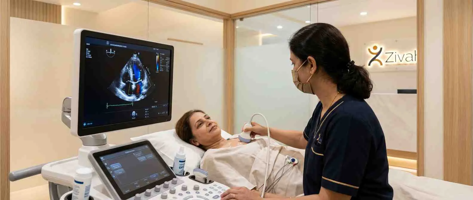
This is what an echocardiogram looks like, start to finish. You lie on your side, with some ECG stickers stuck to your chest. The sonographer doing the scan, who is a certified echocardiographer, puts a bit of gel on and moves a handheld transducer over your chest.
The probe sends sound waves to your heart and picks up the echoes that return. It is painless, involves no radiation and takes about 40 to 60 minutes.
Your Echocardiogram Appointment: Step by Step
| Stage |
What Happens |
Time |
|---|---|---|
| Before |
Change into gown; ECG stickers placed |
~10 min |
| During |
Gel + transducer images the heart |
30–60 min |
| After |
Resume normal activity (TTE) |
Immediate |
Understanding Your Echocardiogram Results
One key thing to keep in mind first: the notes below are a broad background to help you make sense of your scan, not a diagnosis. Always have an echo test checked by a specialist who reviews the results alongside your symptoms, age, and history before any decisions are made.
When the scan ends, it turns into a written echo test report to go with the moving visuals. To you, this seems to be a wall of numbers and terms, but to the trained eye, these details convey a clear tale about how well your heart is working.
Deciphering the Report: What a Normal vs Abnormal Echo Means
Basically, a normal echocardiogram shows chambers of normal size, walls that contract well, and valves that open and close properly. So what does an abnormal echocardiogram mean?
It suggests something that is not quite that picture, perhaps a weaker pump, a leaky valve, or an enlarged chamber. These results on the echocardiography alone are not a diagnosis; they tell your doctor where to examine next.
Primary Measurements in Echocardiography: Ejection Fraction & Output
A handful of critical numbers says a lot. The most important one is called ejection fraction. That’s the percentage of blood your heart pumps out with each beat. About 55–70% is normal. Your report can include mention of cardiac output and valve function. For a thorough breakdown of these parameters, including condition-specific ranges and how each view is read, check the table.
Echo Findings and What They May Suggest (Clinician-Interpreted)
| Echo Finding |
What It May Suggest |
Typical Next Step |
|---|---|---|
| Reduced ejection fraction (below ~55%) |
Heart failure / weak pumping |
Cardiology review |
| Valve leak or narrowing |
Valve disease |
Further assessment |
| Enlarged chambers / thick walls |
Cardiomyopathy, hypertension effect |
Specialist follow-up |
An echocardiogram is interpreted by a clinician in your full clinical context; findings alone are not a diagnosis.
What an Echocardiogram Can and Cannot Detect
An echo is a powerful test, but it is not all-seeing, and knowing where it shines and where it ceases is really valuable. What a cardiac ultrasound reveals:
- The size and shape of your heart
- How hard does he beat each pump
- The extent to which the valves open and close
- High blood pressure is a sign of stress; echoes of hypertension can be seen thickening the walls before symptoms
But it does have its blind spots. An echo can’t directly see the coronary arteries, the tiny channels that nourish the heart muscle. So if there’s a blockage there, you need a different test. And since it’s generally a picture taken when you’re at rest, it might miss problems that only show up when you’re active.
Can an Echocardiogram Detect Heart Failure or a Heart Attack?
Here are two of the most common questions, and the responses depend:
- Heart failure? Yes. An echocardiogram is one of the main ways heart failure is diagnosed and tracked over time by evaluating ejection fraction and heart anatomy.
- Heart attack? Partly. An echo can reveal the muscular damage of a heart attack, but it can’t see the artery that’s blocked to produce it, that takes more imaging.
Beyond the Echo: When Further Cardiac Tests Are Needed
No single test does everything; therefore, an echo generally refers to the next test. Cardiac catheterisation (angiography) directly scans the coronary arteries if a blockage is suspected. If your symptoms occur only with exertion, a stress echocardiogram is meant to detect them. And when blood flow needs a closer look, a Doppler study fills in the details.
Echocardiography Testing at Zivah
We’ve developed an echocardiography test at Zivah that is easy to perform and practical. The typical scan is a non-invasive cardiac ultrasound, performed in the clinic itself, no needles, no radiation, just a probe gently brushed over your chest.
This heart echo scan is not just a one-off test for many of our patients, but part of a wider picture: looking at the mother’s heart during pregnancy, or screening heart health before conception for women with known cardiac issues. You don’t have to run from clinic to clinic or repeat your narrative, because it’s all part of your treatment journey.
The scan is done by a trained sonographer and read by a specialist, so you get a clear diagnostic echo image and an expert assessment of its significance. It’s a non-invasive heart test that is quick, comfortable, and easy to act on, and, as every patient has different demands, cost is discussed during your appointment rather than being set in advance.
Your Care Pathway: From Heart Echo Scan to Cardiology Review
Here’s the drill. Following your scan, a specialist will review your echo test report alongside your symptoms and history, and explain what the results suggest and what happens next, whether that’s reassurance, monitoring, or a further test. Schedule your echocardiography with Zivah today.