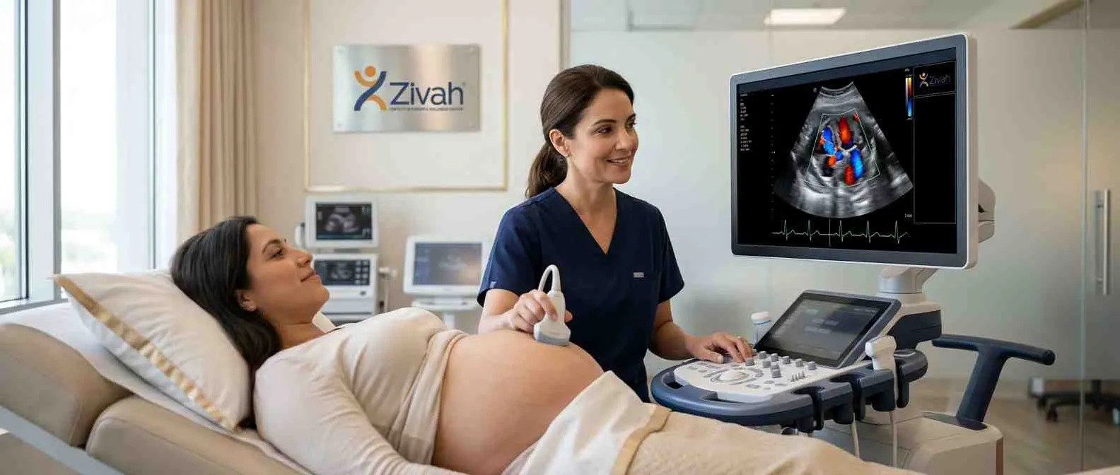
A fetal echocardiography, commonly called a fetal echo or fetal echocardiogram, is a specialist ultrasound that closely examines your baby's heart before birth.
This detailed fetal cardiac ultrasound is more than a basic fetal heart scan and uses special imaging and Doppler to look at the structure of the heart, how well it pumps, the blood flow through the chambers, and the rhythm, giving you a full picture of how your baby's heart is developing and whether it is healthy.
This page explains who needs the test, when it's done, the heart views examined, the conditions it can detect, and how to read your report. Pregnancy care gives you the clearest view of your baby's heart.
What Is Fetal Echocardiography?
A fetal echo is a special heart scan for your unborn baby. Although a oad fetal heart ultrasound provides a rapid overview, this fetal cardiac ultrasound is dedicated to the heart only. It uses 2D imaging with colour and pulsed Doppler to show the heart's anatomy and blood flow.
The Doppler part is important. It can't read electrical signals like a fetal EKG or ECG, but it can indirectly sense the heart's rhythm by monitoring blood flow. (Even a PR-interval estimate, if you want.)
Just to clarify, this checks your baby's heart, not your own. A maternal echocardiography, a unique test that measures the mother's own heart during pregnancy.
Fetal Echo vs Anomaly Scan: Key Differences
So how is this different from your anomaly scan? The TIFFA scan measures the entire fetal body; however, it only provides a basic view of the heart. A fetal echo is a specialised fetal cardiac scan, a far more detailed evaluation that focuses just on congenital heart problem screening. An appointment is normally scheduled if the anomaly scan shows something or if there is a risk factor.
Fetal Echocardiography at a Glance
| Clinical Parameter | Zivah Protocol Detail | Patient Significance |
|---|---|---|
| Study type | Targeted fetal cardiac ultrasound | Heart-focused, not a general scan |
| Timing | 18–24 weeks; early echo 12–16 weeks if high-risk | Best window for clear cardiac views |
| Approach | Transabdominal; transvaginal early or for position | Mostly belly-surface imaging |
| Modality | 2D + colour and pulsed Doppler | Shows structure and blood flow |
| Duration | ~30–60 minutes | Longer, as each view is checked |
| Preparation | Minimal; no full bladder needed | Little to arrange beforehand |
Why Is Fetal Echocardiography Done?
Heart problems are more common than many parents know; congenital heart disease occurs in about 8 to 10 of every 1,000 babies and is the most common congenital disability. The major reason a fetal echo is recommended is that it catches these issues early, when there’s still time to prepare.
A close fetal cardiac assessment does more than check that the heart looks fine. If something is detected, early screening for congenital heart disease in fetuses allows your team to plan the safest possible delivery and arrange specialist treatment from day one, rather than reacting after birth.
What Fetal Echocardiography Helps Your Care Team Plan
A full heart assessment during pregnancy provides your doctors with a good start. The results help them:
- Identify heart problems earlier.
- Choose a hospital properly equipped to care for newborn hearts.
- Consult a children’s heart specialist if necessary.
- Find out if treatment before birth is an option.
- Match the genetic test with the heart results.
Who Needs a Fetal Echocardiogram?
This scan is not needed in every pregnancy. Unlike the anomaly scan, a fetal echo isn't standard for everyone; it's recommended when there's a special need to examine the baby's heart more closely. It's not an "everybody" check, more like targeted heart monitoring for a higher-risk pregnancy.So who is it for? Your doctor may suggest a fetal echo if a fetal cardiac risk assessment turns up something, whether that's your own health, your family history, or an earlier scan. Below, we multiply the triggers into two major categories, maternal and fetal/family.
Maternal Risk Factors for a Fetal Heart Scan
Some are about the health of the mother herself. Fetal echo is often recommended if you have:
- Pre-existing diabetes, particularly if the HbA1c is increased.
- Lupus or SjöSjögren'sth anti-Ro/SSA or anti-La/SSB antibodies that may lead to emyonic heart block.
- A first-trimester infection, such as rubella or CMV.
- Some medications: lithium, certain anti-epileptics or SSRIs.
- IVF or Assisted Reproduction Pregnancy.
Fetal & Family Risk Factors
Other explanations originate from the baby or the family:
- A parent or sibling with a heart defect present at birth.
- Abnormal NT scan or first trimester screen, see NIPT.
- Anomaly Scan (TIFFA) probable chromosomal disease or anomaly.
- A baby with an abnormal heartbeat or twins that share a placenta.
- An echogenic intracardiac focus (EIF) on the anomaly scan.
Fetal Echocardiography: Common Risk Indications
| Risk Category | Specific Indication | Why a Fetal Echo Is Advised |
|---|---|---|
| Maternal health | Pre-pregnancy diabetes, raised HbA1c | Higher chance of cardiac defects |
| Maternal autoimmune | Lupus/Sjögren's, anti-Ro/La antibodies | Risk of fetal heart block |
| Maternal exposure | Certain meds; rubella/CMV infection | Possible effect on heart formation |
| Family history | Parent or sibling with CHD | Inherited cardiac risk |
| Abnormal screening | Abnormal NT scan or NIPT result | Flags need for closer heart check |
| Fetal findings | Extracardiac anomaly, arrhythmia, EIF | Direct cardiac concern raised |
| Pregnancy type | IVF/ART; monochorionic twins | Statistically higher CHD risk |
When Is Fetal Echocardiography Done in Pregnancy?
The time is similar to your other thorough scans. The ideal period is 18 to 24 weeks, when the heart is big enough to see clearly, but there's still time to plan if something needs to be corrected. This usually falls within your 20-week scan stage, but a fetal echo is a far more detailed examination of the heart than that routine check.
In higher-risk situations, an early echo can be performed around 12 to 16 weeks, with a follow-up scan at roughly 20 to 24 weeks to confirm the findings.
Early vs Standard Fetal Echo Timing
Early or first-trimester fetal echo is commonly performed with a transvaginal probe. At about 13 weeks, the heart is too small to be resolved by a belly scan. Fetal heart development offers detailed transabdominal views through the conventional window. If you merely want to check a heartbeat or heart rate, it is the Early Pregnancy Scan and not this test.
What Does Fetal Echocardiography Check? Fetal Heart Anatomy & Views
This is where the scan earns its name. A fetal echo is not just a quick image, but a series of standard fetal heart images that each reveal a different slice of the heart's structure. They had the specialist map the entire organ: the four chambers, the valves between them, the major vessels leaving the heart, and how all these pieces fit together.
The scan looks at the anatomy, but also the rhythm of the heart and how well that pump is working. The goal at this stage is simply to confirm normal fetal heart anatomy; specific conditions are covered later on this page.
Standard Fetal Heart Views Examined
Before examining the heart itself, the specialist confirms the situs, which way round the baby is lying, so left and right are accurately identified. If that is off, the whole map of the heart changes.
From there, the chambers, outflow passages and arches of the heart are swept in turn. No one image is enough. The four-chambered view + the outflow views together catch the majority of important heart abnormalities.
Fetal Circulation, Blood Flow & Cardiac Function
There is more to structure than just the story. The doctor uses colour and pulsed Doppler to examine the blood flow through the baby's heart, making sure it's flowing in the proper direction and that the valves aren't leaky.
It also measures the strength of the heart's pumping and confirms that the cardiac activity is within a normal rhythm, with a heart rate of about 120-160 beats per minute.
Fetal Heart Views & What Each Assesses
| Cardiac View | Structures Visualised | Clinical Purpose |
|---|---|---|
| Situs / abdominal view | Stomach, vessels, left–right orientation | Prerequisite, confirms correct sidedness |
| Four-chamber view | Both atria, ventricles, AV valves | Core screen for chamber defects |
| LVOT | Left ventricle to aorta connection | Checks aortic outflow |
| RVOT | Right ventricle to pulmonary artery | Checks pulmonary outflow |
| Three-vessel / 3VT | Pulmonary artery, aorta, SVC, trachea | Detects great-vessel anomalies |
| Aortic & ductal arch | Aortic arch, ductus arteriosus | Assesses arch formation |
| Bicaval view | SVC and IVC into right atrium | Confirms venous drainage |
Conditions Fetal Echocardiography Can Detect
So what is a fetal echo good for finding? Quite a bit. In the hands of expert personnel, it can identify the most significant cardiac defects: gaps between the chambers, valve difficulties, abnormalities of the main veins, and heart rhythm problems. It can also detect an enlarged fetal heart (cardiomegaly) and indicators that the heart is not pumping as well as it should.
But it’s worth being honest about limits. Some faults are small; others only appear later in pregnancy or after birth; thus, however comforting the scan, not every potential problem can be ignored. The conclusions were largely about structural problems and rhythm.
1. Structural Heart Defects Detected
Structural faults are problems with the way the heart is built. Doctors frequently sort them according to whether they influence oxygen levels.
Acyanotic defects, like a ventricular septal defect (VSD) or atrial septal defect (ASD), are holes between chambers. Tetralogy of Fallot or transposition of the major arteries are cyanotic disorders and impair the delivery of oxygen-rich blood to the body. The table covers these plus coarctation and undeveloped heart syndromes.
2. Fetal Heart Rhythm & Function Abnormalities
Beyond the structure, the scan checks the heart pulse itself. It can pick up a foetal arrhythmia, a heartbeat that is too rapid, too slow or irregular and foetal heart block, which is sometimes linked with the maternal anti-Ro/La antibodies indicated above. This is compared with Doppler timing of the PR interval, while cardiomegaly signals a heart that is under pressure.
How Accurate Is Fetal Echocardiography?
In qualified hands, a fetal echo is very accurate. The four-chamber view alone detects about 60% of significant cardiac abnormalities, and adding outflow-tract images increases that to about 90%. But some problems are not detected until later in pregnancy or after the baby is born.
Congenital Heart Conditions Detectable on Fetal Echo
| Condition | Type | What It Involves |
|---|---|---|
| VSD / ASD | Acyanotic | Hole between heart chambers |
| AV septal defect | Acyanotic | Shared central valve/septal opening |
| Tetralogy of Fallot | Cyanotic | Four combined cardiac abnormalities |
| Transposition (TGA) | Cyanotic | Main arteries connected the wrong way |
| HLHS / HRHS | Cyanotic | One side of the heart underdeveloped |
| Coarctation / pulmonary stenosis | Structural | Narrowing of a major vessel/valve |
| Arrhythmia & heart block | Rhythm | Abnormal or blocked heartbeat |
How Is Fetal Echocardiography Performed?
From your side, this just feels like any other pregnancy ultrasound. You lie down, they apply some gel to your tummy, and the specialist moves a transducer across your skin to capture an image of your heart from multiple angles.
The scan normally takes 30 to 60 minutes because each view is carefully captured, and it may take longer if the baby is in a difficult position. The photos are then reviewed by a fetal medicine or cardiology professional. What can influence the picture? How to prepare. Is it safe? These are covered in the parts below.
How to Prepare for a Fetal Echocardiogram
Hardly anything to prepare in advance. Wear loose clothes that you can pull away from your bump. No need to fast or fill your bladder. Please ing your referral letter and any previous scan reports. Sometimes a transvaginal view is used, usually early in pregnancy or if the position of the baby makes the heart difficult to see.
Acoustic Window & Diagnostic Limits
Sometimes the picture is not as clear as you'd like; that's down to physics, not anything wrong. The ultrasound has to pass through tissue to reach the heart; thus, a thicker abdominal wall, an anterior placenta, low amniotic fluid, or a face-down baby might all compromise the view. When this happens, a short repeat scan is often arranged to complete the missing images.
Is Fetal Echocardiography Safe During Pregnancy?
Yes, very safe. A fetal echo uses just sound waves, no radiation, nothing intrusive, hence no known risks to you or your baby. This is how ultrasound has been utilised in pregnancy for decades.
Worried about your baby's heart? Referred for a fetal echo? Book your prenatal echocardiogram at Zivah for detailed heart imaging and specialist assessment.
Understanding Your Fetal Echocardiography Report
After the scan, you will be given a written report, usually reviewed by a fetal-medicine or paediatric cardiologist. It can sound like a series of weird words, but the concept is simple: it discusses the structure of the heart, the blood flow through it, its rhythm and how well it pumps, then ends with a summary.
A normal result indicates nothing turned up in any of them. However, one honest note: a baby's circulation works differently from a newborn's, thus a few small findings can only be validated after birth.
What a Normal Fetal Echo Report Means
A normal fetal echocardiogram is reassuring; it means the heart's shape, flow, and rhythm all appear as predicted for this time. Remember this is a strong indicator of heart health, not a cast-iron guarantee, as extremely small or late-developing abnormalities might remain unnoticed until delivery.
Understanding Your Fetal Echo Report
| Report Component | What It Measures / Describes | Clinical Implication |
|---|---|---|
| Cardiac structure | Chambers, valves, great vessels | Confirms the heart is correctly formed |
| Blood flow | Doppler flow direction and valve function | Checks circulation through the heart |
| Rhythm & rate | Heartbeat pattern and speed | Flags arrhythmia or heart block |
| Cardiac function | Pumping strength (MPI) | Shows how well the heart works |
| Summary & recommendation | Overall conclusion and next steps | States what, if anything, follows |
What Happens If a Fetal Heart Abnormality Is Found?
If the scan reveals a heart problem, it doesn't mean a choice has to be made straight away. First, there is clarity, knowing exactly what has been observed and how important it is.
Your professional will explain the findings clearly and provide a clear plan. That depends on the type of fetal heart defect. Some are small and just monitored; others need a closer review from a bigger team. Either way, it now buys time to prepare properly, as the next part sets out.
Next Steps & Multidisciplinary Care
Depending on what is found, your care may include:
- Repeat or serial echos to monitor the heart over time.
- A pediatric cardiology consult and your fetal-medicine (MFM) team.
- Optional Amniocentesis if testing for a genetic cause.
- Genetic Counselling to discuss what a result means.
Is fetal cardiac intervention recommended? Many small VSDs close on their own before or after birth, thankfully.
Why Choose Zivah for Fetal Echocardiography?
The actual question is who is reading the scan and how carefully when you are looking for a fetal echocardiogram near me. Zivah has a highly skilled team of fetal medicine and cardiology specialists who evaluate your baby’s heart using modern 2D and colour Doppler imaging for detailed cardiac testing.
Each scan is done using a systematic view-by-view methodology, and your report is not merely passed over but is analysed by an expert. If a discovery requires more support, counselling, and follow-up, they are organised under one roof so that you won't be sent between clinics for the next step.
Expert Fetal Cardiac Imaging at Zivah
It comes down to experienced personnel and capable equipment for accurate results. Zivah is a specialist foetal cardiac imaging clinic. We pair skilled sonographers with fetal-medicine and cardiology input on high-resolution Doppler devices designed for advanced cardiac screening.