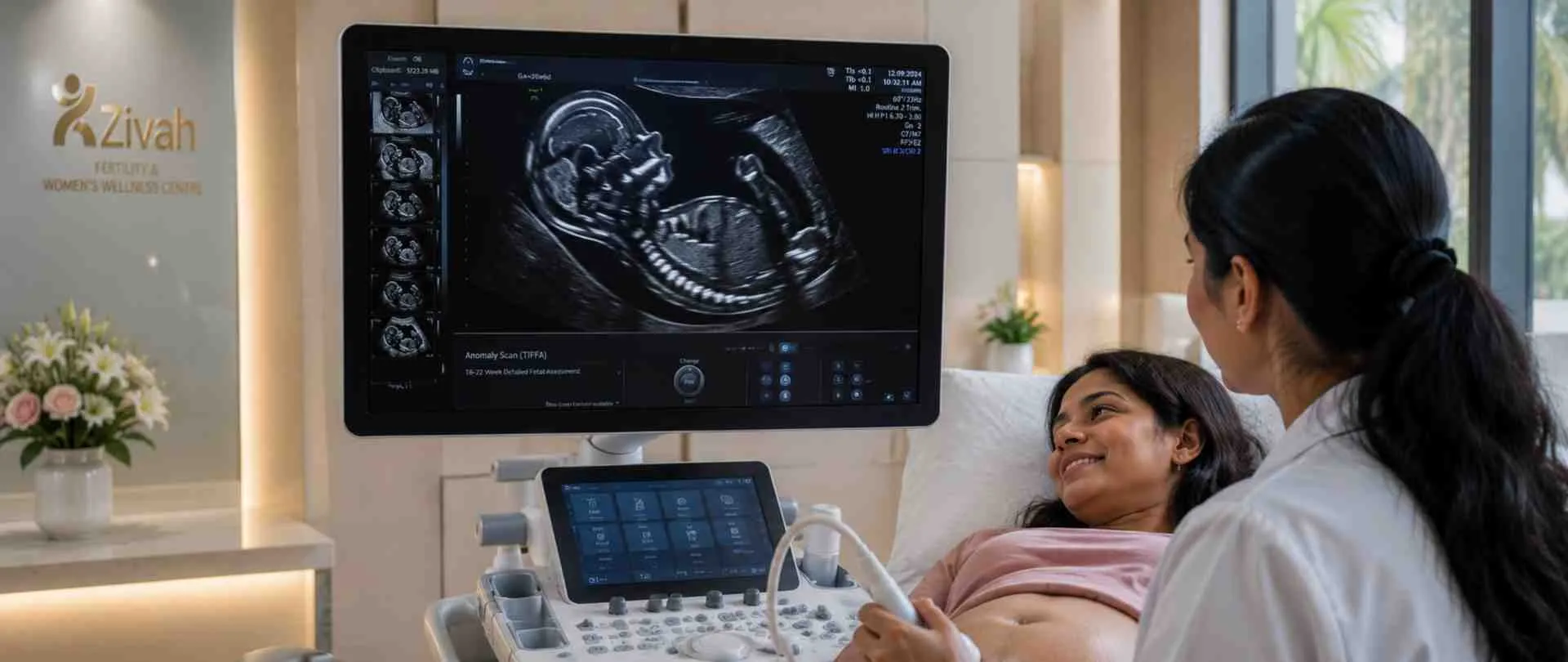
Anomaly scan, also known as TIFFA scan or Level 2 ultrasound, is a detailed second-trimester diagnostic scan that uses targeted imaging for fetal anomalies.
The fetal anomaly scan is done between 18 and 22 weeks of pregnancy to check your baby’s developing anatomy from head to toe the brain, face, spine, heart, kidneys and limbs to make sure each organ system is developing as planned at this stage.
This page explains when your scan is scheduled, which anatomical structures the specialist will be looking for, how you should prepare, and what each report result means for the rest of your pregnancy.It is still the most important structural progress your baby makes in routine prenatal care.
What Is the Anomaly Scan (TIFFA Scan)?
At about the fifth month, the specialist will closely examine the formation of your baby’s organs one by one. This is the core of this scan: a detailed second-trimester ultrasound that charts the baby’s full anatomy, hence why it is also known as a fetal anatomy scan or morphology scan. Its concern is just structural.
It examines the development of organs and body parts, but it is not a standalone chromosomal test; a clear result will not substitute for genetic screening such as NIPT.
Let’s clear up a common mix-up: an anomaly scan and a 3D scan are not the same. TIFFA is a diagnostic 2D anatomy survey read by a fetal medical specialist, not the optional 3D/4D “baby photo” imaging done. Our blog has much more information about the specific situations our scan can detect.
What Does "TIFFA" Mean? (Targeted Imaging for Fetal Anomalies)
The acronym is Targeted Imaging for Fetal Anomalies, targeted imaging to find structural differences in the baby. In common use, it is just another name for the anomaly scan.
Why It's Called a Level 2 Ultrasound
Pregnancy ultrasounds are rated on their level of detail. Growth and dating are a Level I scan, the basic in-depth morphology scan, where you look at each anatomical feature up close. It is a Level II study, and this scan is at that level.
Anomaly Scan (TIFFA) at a Glance
| Clinical Parameter |
Zivah Protocol Detail |
Patient Significance |
|---|---|---|
| Scan type |
Level II anatomy ultrasound |
Detailed structural survey, not a routine growth check |
| Timing |
18–22 weeks |
Optimal window for clear organ views |
| Approach |
Transabdominal; TVS only for cervix or unclear views |
Mostly belly-surface; internal only when required |
| Duration |
30–45 minutes |
Longer than a standard scan — every structure is checked |
| Preparation |
Minimal |
No fasting; light bladder filling may be advised |
| Report |
Usually same day |
Results reviewed without a long wait |
When Is the TIFFA Scan Done in Pregnancy?
The timing for this scan is more important than for practically any other scan throughout pregnancy. It’s done in the second trimester, between 18 and 22 weeks (that’s your 5th month), and most specialists like to schedule it in the smaller 18–20-week window.
By this point, the baby is large enough for each organ to be seen clearly, but still small enough to fit easily in the frame. It is that balance that makes the 20 week pregnancy scan the typical checkpoint for a thorough anatomy survey.
If your dates are a bit off or the first pass does not capture all the views, you can rescan within the same time window without losing accuracy. Booking your second-trimester anomaly scan early in the range just gives you more room for follow-up if anything needs a closer look.
Why 18-22 Weeks Is the Ideal Window

There are some practical, clinical reasons why the best time for an anomaly scan is in this range:
- The main organs: heart, brain, spine and kidneys are fully formed and large enough to be pictured in great detail.
- The baby is bathed with enough amniotic fluid for crisp, undistorted images.
- The baby still moves freely so that the specialist can get all the angles the survey needs.
- This provides a comfortable buffer for a repeat scan, other tests or specialist assessment if needed.
What If the Scan Is Done Later?
It is this combination that makes the 20-week scan the standard for a full structural assessment. And sometimes an anomaly scan late beyond 22 weeks is still achievable and often fully legible. The sooner, the better; opinions tend to be clearer, and there’s more time for any follow-up the report may propose.
Why Is the Anomaly Scan Important in Pregnancy?
And this is the most complete structural check your pregnancy gets by the second trimester. It is highly specific in its intent: to examine the development of all major organs and body parts and to identify any anatomical abnormality while it can still be addressed. The value is in early knowledge.
Early identification of a structural problem enables your care team to prepare rather than react, scheduling confirmatory tests, specialist consultations, or a customised delivery plan well in advance.
A normal scan, on the other hand, shows a definite structural baseline in your records. That combination of detection and planning is what gives the TIFFA scan its weight in ordinary prenatal care.
What the TIFFA Scan Helps Your Care Team Plan
A clear picture at this point gives your doctors hard data to work with. In practice, the scan supports:
- Early detection of fetal anomalies – when variances in structure are at their broadest and with it management choices.
- Delivery and birth-setting planning – picking the right hospital or unit in case you need expert newborn care.
- Specialist input – if a finding justifies it, referral to fetal medicine, cardiology or genetics.
- Follow-up testing – timely sequencing of any confirming scans or diagnostic testing.
These, together, make up the basis for the rest of your prenatal care strategy.
What Does the TIFFA Scan Check? (Fetal Anatomy Examined)
This fetal anatomy scan isn't a quick look; it's a detailed, step-by-step look. The doctor will undertake a head-to-toe examination of your baby's body and check each of the major organs as she goes. They also look at the structures surrounding the baby, such as the placenta and fluid, which help in keeping the pregnancy on track.
Please note: The sex of the fetus is neither determined nor disclosed in this scan under the PCPNDT Act, 1994.
This is a fetal organ screening ultrasound; thus, the focus is primarily on anatomy, how each structure is created, sized and positioned. In the examination, both the infant's development and its environment, the placenta and amniotic fluid are assessed.

Head-to-Toe Fetal Structural Assessment
This section of the fetal development scan goes through a checklist from top to bottom:
- Brain and skull: form, midline, cerebral ventricles Face and lips: profile, upper lip and eye-holes.
- Spine: Straightness and skin covering down the whole length.
- The heart: four chambers, valves, and major outflow channels.
- Chest and Lungs: Symmetry and Diaphragm.
- Abdomen and stomach: wall, bubble of stomach and intestine.
- Kidneys and bladder: both kidneys and urine flow.
- Limbs: arms, legs, hands, and feet, with bone lengths measured.
This scan takes a close look at the heart, but there is a limit to the detail it can give. A more detailed assessment of the heart, if needed, is done with a second test called a fetal echocardiogram.
Placenta, Amniotic Fluid & Umbilical Cord Assessment
A baby is as important as the baby’s environment. The three supporting structures are reviewed as:
- Placenta - its position is identified, especially if low-lying or covering the cervix, which helps in planning delivery.
- Amniotic fluid - the volume of amniotic fluid is assessed since too little or too much may indicate a problem.
- Umbilical cord - Check the number of vessels and the connection to the placenta.
Each of them tells the staff how well the pregnancy is being supported.
Fetal Anatomy Examined on the Anomaly Scan
| Anatomical Region |
Target Structural Elements Checked |
Clinical Rationale |
|---|---|---|
| Head & brain |
Skull shape, midline, cerebral ventricles |
Confirms brain structure and ventricle size |
| Face & spine |
Lip/profile, vertebral alignment, skin cover |
Screens facial and spinal formation |
| Heart & chest |
Four chambers, valves, outflow tracts |
Core cardiac and lung-field check |
| Abdomen & kidneys |
Stomach, bowel, renal structures, bladder |
Assesses digestive and urinary organs |
| Limbs |
Arm/leg bones, hands, feet |
Confirms limb length and completeness |
| Placenta & fluid |
Placental position, amniotic fluid volume, cord |
Gauges the baby's support environment |
What Conditions Can the Anomaly Scan Detect?
This scan is best for looking at how your baby is built, and that is where it works hardest. It picks up the most serious congenital abnormalities of the brain, spine, heart, kidneys and limbs, which is why it is the principal structural anomaly check in pregnancy. But it still cannot fulfil all promises.
A normal scan reduces the risk of serious disease, but it cannot rule out all conditions. Some are too small to see, and others only appear later in pregnancy. The table below shows how frequently the scan detects distinct conditions.
Why the Anomaly Scan Is Not a Chromosomal Test
The scan looks at structure, not at chromosomes. It can look for physical signs that sometimes accompany a chromosomal problem, but it cannot directly examine the chromosomes.
This is most evident with Down syndrome; only around half of the cases are detected by the anomaly scan, far lower than for diseases like anencephaly. It supplements, but never replaces, specialised screening. See NIPT and First Trimester Screening for chromosomal testing.
What Are Soft Markers on an Anomaly Scan?
Sometimes a report will say "soft marker", and that one term causes a lot of anxiety. Here's what it truly means. Soft markers are subtle signs that are not problems themselves, like an echogenic intracardiac focus (a bright point in the heart), a choroid plexus cyst in the brain, echogenic bowel, mild pyelectasis (slightly full kidneys), a short thigh bone, or a thicker neck fold.
Most are normal and go away on their own. Now and then, one or a few, in combination, merit closer study. Then your team may offer NIPT, Amniocentesis, and Genetic Counselling.
Anomaly Scan Detection Rates by Condition
| Condition |
Type |
Approx. Detection on Anomaly Scan |
|---|---|---|
| Anencephaly |
Structural |
~98% |
| Open spina bifida |
Structural |
~90% |
| Edwards' syndrome (T18) |
Structural features |
~95% |
| Patau's syndrome (T13) |
Structural features |
~95% |
| Down syndrome (T21) |
Chromosomal |
~50% |
| Congenital heart disease |
Structural |
50% |
How Is the TIFFA Scan Performed?
This examination is nothing hard on your side, it's a regular ultrasound, just slower and more detailed. You lie back as the sonologist moves a tool over your belly, and there is your baby, alive on the screen. Below, we walk you through the process, what to bring, and whether it's safe.

The Step-by-Step Anomaly Scan Procedure
A small amount of warm gel is applied to your abdomen, and the sonographer moves a handheld transducer over your skin to obtain real-time images. They then carefully work through each structure, pausing to measure and save the important views before completing the study of the placenta and cord.
Fetal Position & the Acoustic Window: Why a Repeat May Be Needed
Sometimes the baby just won't cooperate. A face-down or breech presentation, plenty of movement, or denser body tissue may mask some images, such as the outputs of the heart or the profile. If so, you may be invited to return for a short top-up scan that day or week. Routine quality control, not a warning indicator, is getting called back for an anomaly scan.
How to Prepare for Your TIFFA Scan
Not much is required beforehand:
- Wear loose clothes you can pull away from your tummy.
- No fasting. Eat and drink regularly.
- Having a somewhat full bladder helps with the early images, so take a little water on board before you arrive.
- Bring your prescription and previous scan reports with you.
The specialist may even include a fast transvaginal examination to determine cervical length and a preterm-birth risk analysis.
Is the Anomaly Scan Safe During Pregnancy? Yes. Ultrasound uses sound waves, not radiation, so it's non-invasive and poses no known adverse effects for you or your baby. It's also been safely used throughout pregnancy for decades.
Understanding Your TIFFA Scan Report
When the scan is completed, you will receive a written anomaly scan report, usually the same day. To begin with, it may look like a wall of numbers and medical terms, but the layout is simple: your baby's measures, the look of each organ, the placenta, fluid and position, and a summary at the end. Below, we explain what a typical result means and what each portion of the TIFFA scan report is telling you.
What a Normal TIFFA Scan Report Means
A "normal" result means that the doctor looked at every structure they could see and didn't see anything abnormal - your kid is growing as they should. You may also see "low risk for structural anomaly".
Good news: Just remember, the scan reads structure, not everything; thus, a normal anomaly scan minimises the chance of a problem without being an absolute guarantee.
What the Anomaly Scan Report Includes
Most reports have the same sections: fetal biometry (measurements of the head, abdomen, and femur that indicate growth), an anatomy scan of each organ, a mention of the placenta and fluid, the baby's position, and a final summary of any next steps. What each portion signals is shown in the table below.
Understanding Your TIFFA Scan Report
| Report Component |
What It Measures / Describes |
Clinical Implication |
|---|---|---|
| Fetal biometry |
Head, abdomen, and femur measurements |
Confirms growth matches gestational age |
| Fetal anatomy |
Organ-by-organ structural findings |
Flags or clears any structural concern |
| Placenta & amniotic fluid |
Placental position, fluid volume |
Checks the baby's support environment |
| Position |
How the baby is lying |
Notes views affected; may explain a recall |
| Summary & recommendation |
Overall conclusion and next steps |
Tells you what, if anything, happens next |
What Happens If an Abnormality Is Found?
First, an “abnormal” or confusing discovery doesn’t necessarily signal something is significantly wrong; many turn out to be benign, transient or only need a second look. Should anything be flagged on your TIFFA scan report, the professional will explain what they saw in straightforward words, how certain they are and the reasonable next action.
This can be a repeat scan in a couple of weeks, or a referral for closer assessment. The goal here is understanding, not answers; each stage simply confirms what’s happening so your team can prepare.
Next Steps After an Abnormal Anomaly Scan
Depending on what is found, future steps usually include one or more of the following:
- Repeat scan or confirmatory scan - a closer or later inspection to validate the finding.
- Fetal medicine referral - more detailed image assessment by an MFM specialist.
- Diagnostic testing - Optional tests, such as amniocentesis, if needed.
- Genetic counselling - a chat to help comprehend what a result means.

Your team explains every choice before making a decision.
Is the TIFFA Scan Necessary in Pregnancy?
The honest answer is this. There is no legal requirement to have any scan, but the anomaly scan is strongly advised for all pregnancies as part of routine antenatal care. Most obstetricians consider it an important checkpoint, not an optional extra.
The explanation is simple: it’s the only time in pregnancy when your baby’s whole structure can be examined in full detail, early enough to act on anything identified.
Not doing so means missing that window. That said, the final decision is yours, in partnership with your doctor. If you have questions about whether it’s right for you, your obstetrician is best placed to discuss things through.
Who Should Have an Anomaly Scan?
Short answer: anybody. The scan is available to all pregnant women, regardless of age or history. But it’s more important in a high-risk pregnancy, for example, if you’re over 35, have a family history of birth abnormalities, had a baby with an anomaly before, or had an abnormal result on an earlier screening test. Here, the amount of detail it provides is very valuable.
Why Choose Zivah for Your Anomaly Scan (TIFFA Scan)?
The biggest thing is who reads the images and how thoroughly, if you're looking for an anomaly scan near me. At Zivah, your TIFFA scan will be done and analysed by the expertise of both fetal medicine and radiology, on the latest ultrasound machines for precise fetal imaging.
And every scan is careful, structure by structure, so nothing is missed. You also get clear report interpretation and, if needed, counselling and follow-up, all under one roof, not put off to the next stage.
Expert Fetal Imaging at Zivah
There is a trained eye behind every correct scan. Zivah is a dedicated fetal anomaly scan centre where all prenatal ultrasound scans are performed by professional sonologists with fetal-medicine input, on high-resolution equipment suitable for advanced fetal screening. It is the combination of competent readers and capable imaging that makes a complete anatomy survey reliable.